
Research Tip for the Evaluation of Dysferlin Localization in Skeletal Muscle Sections
Dysferlin is a single pass transmembrane protein that is highly expressed in skeletal and cardiac muscle. Historically, dysferlin localization has been shown to be localized only at the sarcolemma of muscle fibers (see Figure S2 below). However, dysferlin’s location within skeletal muscle has evolved with studies showing that while dysferlin is present on the sarcolemma, it is much more abundant in intracellular membranes of skeletal muscle such as in the reticulum of the sarcoplasm and in the transverse tubules (T-tubules) (see Figure 1 below).
Given the diversity in dysferlin protein localization, we highly recommend evaluating dysferlin expression in both cross sections and longitudinal sections in order to get a full picture of where dysferlin is localized and/or impacted in the experimental state being evaluated.
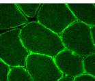 Figure S2 from Yasa et al. ActaNeuropatholCommun. 2003 Jan 18; 11:15
![]()
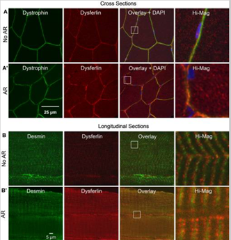 Figure 1 from
 ![]()
Roche et al has shown that the visualization of intracellular staining, especially in cross section, often requires the use of antigen retrieval (AR) methods. In cross section, AR enhances labeling of dysferlin (Fig 1, red labeling), at the intracellular reticulum, but doesn’t change the staining for dystrophin (Fig 1, green labeling). While in longitudinal sections without AR, dysferlin appears as puncta adjacent to the Z-disks. After AR, it is a clear doublet flanking the Z-disks.
In addition, researchers need to consider the following factors that impact the visualization of dysferlin localization when planning their experiments and analysis:
1. Optimization of labeling techniques with antibodies are often needed to properly visualize dysferlin on muscle sections.
2. Antigen retrieval methods are helpful to visualize the complete breadth of dysferlin localization.
3. Proper fixation of muscle is important for immunolocalization of dysferlin in both longitudinal and cross sections.
4. There is potential for background issues depending on the dysferlin antibody used and the species being evaluated. For example, the NCL-Hamlet antibody is a mouse monoclonal antibody which can sometimes cause background issues when staining mouse tissues.
The Jain Foundation identifies and helps develop essential tips and tools for dysferlin research that can be found at the links below. Many of these resources were created through JF funded projects and can be requested by sending email to: admin@jain-foundation.org.


