ACCESS RESOURCES
Animal Models
ACCESS RESOURCES
Animal Models
Home ❯ Research ❯ Access Resources ❯ Research Tools ❯ Animal models
Animal Models
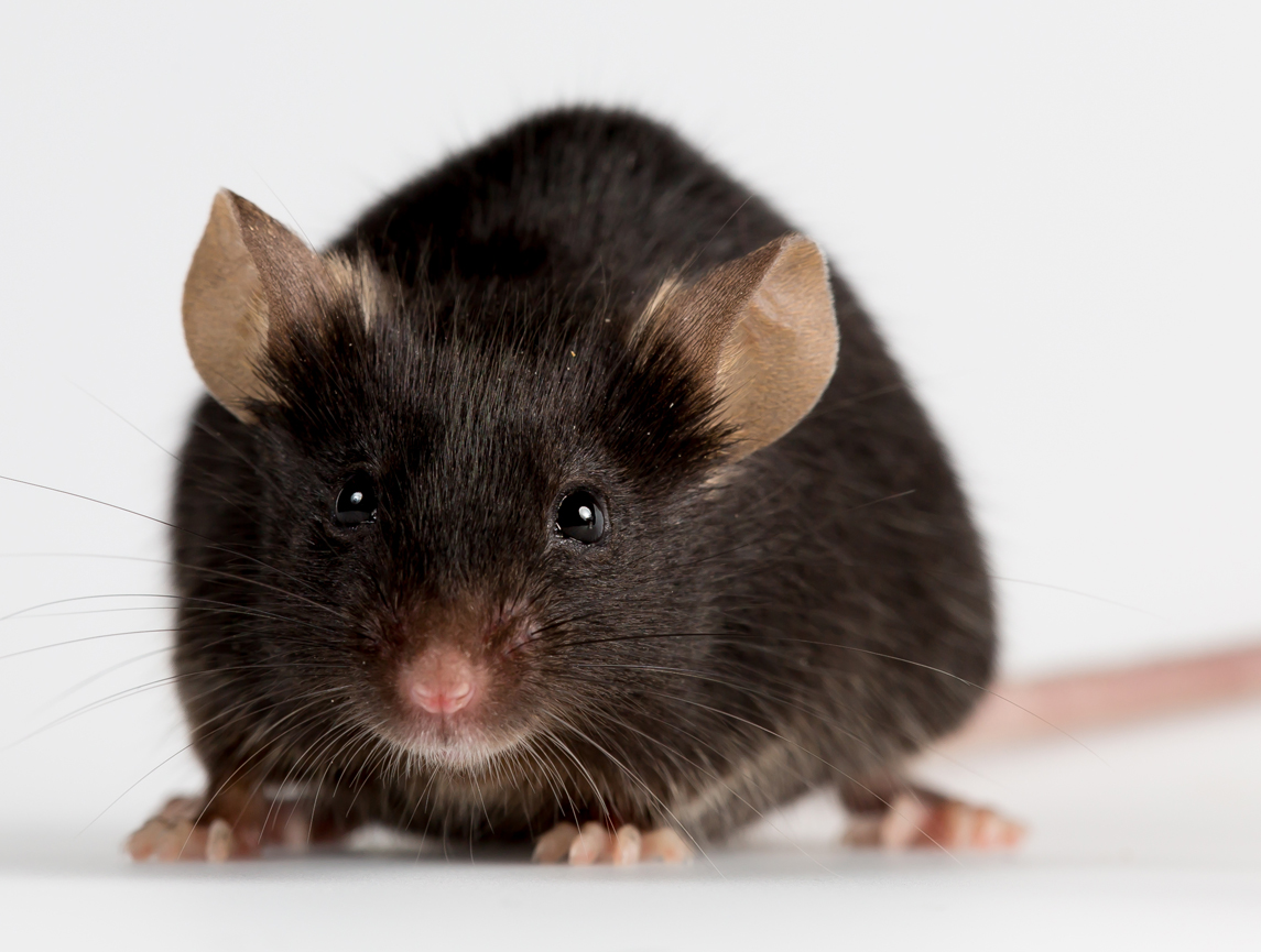
Below is a list of dysferlin deficient mouse lines that are available, with the Bla/J being the most commonly used and recommended for most applications. The majority of the mouse models are available from The Jackson Laboratory and the list includes information on the strain and mouse characteristics. Please note that the Bla/J line is officially cryopreserved by JAX, but the Jain Foundation maintains a private live colony of the Bla/J line for public use. If you are interested in obtaining Bla/J mice contact the Jain Foundation at admin@jain-foundation.org. There are also various zebrafish lines that have been created that are deficient in various muscle related genes, including dysferlin.

Bla/J mice (B6.A-Dysfprmd/GeneJ)
Availability:
The Jackson Laboratory
Stock number: 012767
Strain information: Click Here
Phone: 800-422-MICE (800-422-6423)
610 Main Street, Bar Harbor, Maine 04609 USA
Please note that this strain has been cryopreserved by the Jackson Laboratories, but the Jain Foundation maintains a private colony of these mice for public use. To obtain these mice, please contact the Jain Foundation:
Jain Foundation Inc.
9706 Fourth Avenue NE, Suite 101
Seattle, Washington 98115
Phone: 425-882-1492
Fax: 425-882-1050
Email: admin@jain-foundation.org
Development:
In these mice the progressive muscular dystrophy (prmd) allele from the A/J inbred strain is crossed onto the C57BL/6 genetic background. The cross was performed in the laboratory of Dr. Isabelle Richard at Genethon and the backcross generation reached N8. In collaboration with the Jain Foundation, Dr. Richard donated the strain to The Jackson Laboratory in 2010. Upon arrival, mice were bred to C57BL/6J for at least 2 generations to establish the colony.
Mutation:
An ETn retrotransposon (5-6kb) is inserted in intron 4 of the dysferlin gene.
Symptoms:
Disease onset is observed by 2 months and is characterized by the presence of centronucleated fibers and areas of inflammation. As seen with the original background A/J, mice homozygous for the prmd allele on the C57BL/6J background display an increasing number of centronucleated fibers and impairment in the majority of muscles by 4 months of age. In order of severity, the most affected muscles are psoas, quadriceps femoris, tibialis anterior, and gastrocnemius. Mice exhibit a decreased membrane repair capacity following laser wounding experiments. In an open space assay, mice cover less distance and are less active than wild-type. Mice that are homozygous for this allele are viable, fertile and normal in size.
Comparison with other disease strains:
Disease progression is similar to A/J: slightly slower than in SJL/J, Dysf-/- (Campbell), Dysf-/- (Brown), and C57BL/10.SJL mice. As in both SJL/J and Dysf-/- (Brown) mice, proximal muscles are more severely affected than distal muscles. As in Dysf-/- (Brown) mice, abdominal muscles are also affected.
Control strain(s):
C57BL/6 (for homozygotes); littermates (for heterozygotes).
References:
Lostal W; Bartoli M; Bourg N; Roudaut C; Bentaib A; Miyake K; Guerchet N; Fougerousse F; McNeil P; Richard I. 2010. Efficient recovery of dysferlin deficiency by dual adeno-associated vector-mediated gene transfer. Hum Mol Genet 19(10):1897-907.
- PMID: 20154340
A/J
Availability:
The Jackson Laboratory
Stock number: 000646
Strain information: http://jaxmice.jax.org/strain/000646.html
Phone: 800-422-MICE (800-422-6423)
610 Main Street, Bar Harbor, Maine 04609 USA
Development:
This inbred strain contains a naturally-occurring dysferlin mutation and was originally developed from a cross between a Cold Spring Harbor albino and a Bagg albino.
Mutation:
An ETn retrotransposon (5-6kb) is inserted in intron 4 of the dysferlin gene.
Symptoms:
Histological evidence of dystrophy is not seen until 4-5 months of age and muscle weakness progresses slowly. Abdominal muscles are most severely affected, followed by proximal muscles and then distal muscles. The mice also have other symptoms, including a high incidence of lung adenomas and mammary adenocarcenomas. They are homozygous for an age related hearing loss allele (Cdh23 gene) and for hemolytic complement deficiency (C5, Hc gene).
Comparison with other disease strains:
Disease progression is slightly slower than in SJL/J, Dysf-/- (Campbell), Dysf-/- (Brown), and C57BL/10.SJL mice. As in both SJL/J and Dysf-/- (Brown) mice, proximal muscles are more severely affected than distal muscles. As in Dysf-/- (Brown) mice, abdominal muscles are also affected.
Control strain(s):
A/HeJ, A/WySnJ
References:
Ho M, et al. 2004. Disruption of muscle membrane and phenotype divergence in two novel mouse models of dysferlin deficiency. Human Molecular Genetics 13:1999-2010.
C57BL/6J-Chr6A/J/NaJ mice
Availability:
The Jackson Laboratory
Stock number: 004384
Strain information: http://jaxmice.jax.org/strain/004384.html
Phone: 800-422-MICE (800-422-6423)
610 Main Street, Bar Harbor, Maine 04609 USA
Development:
A chromosome substitution or consomic strain with one of its chromosomes replaced by the homologous chromosome of another inbred strain. The C57BL/6J and A/J strains in this set were chosen because they differ in their susceptibility to diseases such as arthritis, asthma, atherosclerosis, cancer, several infectious diseases, inflammatory responses, and physiological, behavioral and sensory phenotypes. This particular strain has chr 6 from the A/J line with an ETn retrotransposon (5-6kb) inserted in intron 4 of the dysferlin gene, while the remaining chromosomes are from C57BL/6J.
Mutation:
An ETn retrotransposon (5-6kb) is inserted in intron 4 of the dysferlin gene.
Symptoms:
Very similar to A/J.
Control strain(s):
C57BL/6J
References:
Nadeau JH; Singer JB; Matin A; Lander ES. 2000. Analysing complex genetic traits with chromosome substitution strains. Nat Genet 24(3):221-5.
SJL/J mice
Availability:
The Jackson Laboratory
Stock number: 000686
Strain information: http://jaxmice.jax.org/strain/000686.html
Phone: 800-422-MICE (800-422-6423)
610 Main Street, Bar Harbor, Maine 04609 USA
Development:
This inbred strain contains a naturally-occurring dysferlin mutation and was originally developed from three different sources of Swiss Webster mice.
Mutation:
Exon 45 (171 base pairs, aa1628-1685) is deleted in dysferlin mRNA, due to a mutation in the 3’ splice junction. This deletion removes part of the fifth C2 domain (C2E) of the protein.
Symptoms:
Mild myopathic lesions can be detected histologically around 1 month of age, with active myopathy by 6-8 months that primarily affects the proximal muscle groups and manifests as progressive muscle weakness. Muscular atrophy begins at 10 months, and by 16 months half of the muscle fibers are replaced by fat. There is dispute about whether young SJL mice (1-2 months) are stronger or weaker than control mice. The mice also have a high incidence of lymphoma, increased susceptibility to autoimmune diseases and viral infections, and extreme aggression in males, none of which are thought to be associated with dysferlin deficiency. They are also homozygous for a retinal degeneration allele (Pde6b gene).
Comparison with other disease strains:
Disease progression is similar to Dysf-/- (Campbell), Dysf-/- (Brown), and C57BL/10.SJL mice, and faster than in A/J mice. As in both A/J and Dysf-/- (Brown) mice, proximal muscles are more severely affected than distal muscles. The other symptoms, however, are not shared with the other dysferlin-deficient mice and are likely due to complicating genetic features of this strain.
Control strain(s):
No close matches
References:
Weller AH, et al. 1997. Spontaneous myopathy in the SJL/J mouse: pathology and strength loss. Muscle & Nerve 20:72-80.
Bittner RE, et al. 1999. Dysferlin deletion in SJL mice (SJL-Dysf) defines a natural model for limb girdle muscular dystrophy 2B. Nature Genetics 23:141-142.
Vafiadaki E, et al. 2001. Cloning of the mouse dysferlin gene and genomic characterization of the SJL-Dysf mutation. Neuroreport 12:625-629.
NOD.Cg-Rag1tm1Mom Dysfprmd Il2rgtm1Wjl/McalJ
Availability:
Jackson Laboratories stock #029663
Development:
The BlAJ/NRG mutant mice were created by Michele Calos and are an immune-deficient model of dysferlinopathy (also referred to as LGMD2B, LGMDR2, or Miyoshi Myopathy type 1). These mice are deficient in DYSF, Rag1, and IL2rg. The dysferlin mutation is the same retrotransposon insertion that is found in the Bla/J and A/J lines.
References:
- Lostal W; Bartoli M; Bourg N; Roudaut C; Bentaib A; Miyake K; Guerchet N; Fougerousse F; McNeil P; Richard I. 2010. Efficient recovery of dysferlin deficiency by dual adeno-associated vector-mediated gene transfer. Hum Mol Genet 19(10):1897-907; PubMed: 20154340MGI: J:158933
- Ma J; Pichavant C; du Bois H; Bhakta M; Calos MP. 2017. DNA-Mediated Gene Therapy in a Mouse Model of Limb Girdle Muscular Dystrophy 2B. Mol Ther Methods Clin Dev 7:123-131; PubMed: 29159199MGI: J:260166
- Pearson T; Shultz LD; Miller D; King M; Laning J; Fodor W; Cuthbert A; Burzenski L; Gott B; Lyons B; Foreman O; Rossini AA; Greiner DL. 2008. Non-obese diabetic-recombination activating gene-1 (NOD-Rag1 null) interleukin (IL)-2 receptor common gamma chain (IL2r gamma null) null mice: a radioresistant model for human lymphohaematopoietic engraftment. Clin Exp Immunol 154(2):270-84; PubMed: 18785974MGI: J:140388
SCID/BLAJ mice (B6.Cg-Dysfprmd Prkdcscid/J
Availability:
Jackson Laboratories stock #017917
Development:
The first SCID/BLAJ mouse model of dysferlinopathy was created by Dr. Ivan Torrente by backcrossing dysferlin deficient BLA/J mice onto the SCID strain while selecting for the retrotransposon insertion in intron 4 of the dysferlin gene, as well as the absence of specific B- and T- lymphocyte markers (CD4, CD8, and CD19) in cells isolated from peripheral blood. The result is a dysferlin deficient animal model that is also deficient in B and T lymphocytes, and thus more amenable to transplantation studies. Dr. Torrente made the mouse available to the research community in a private colony through the Jain Foundation, but due to low usage, the colony was cryopreserved.
The Jackson Laboratories created a second strain of SCID/BLAJ mice in their own laboratories in 2012, which are currently available in live repository.
Mutation:
An ETn retrotransposon (5-6kb) is inserted in intron 4 of the dysferlin gene.
References:
Farini A, Sitzia C, Navarro C, D’Antona G, Belicchi M, Parolini D, Del Fraro G, Razini P, Bottinelli R, Meregalli M, Torrente Y. 2012. Absence of T and B lymphocytes modulates dystrophic features in dysferlin deficient animal models. Exp. Cell Res. March 23.
Diaz-Manera J, Touvier T, Dellavalle A, Tonlorenzi, R, Tedesco FS, Messina G, Meregalli M, Navaro C, Perani L, Bonfani C, Illa I, Torrente Y, Cossu G. 2010. Partial dysferlin reconstitution by adult murine mesoangioblasts is sufficient for full recovery in a murine model of dysferlinopathy. Cell Death Dis. Aug 5:1e61
Dysf-/- mice (B6.129-Dysftm1Kcam/J)
Availability:
The Jackson Laboratory
Stock number: 013149
Strain information: http://jaxmice.jax.org/strain/013149.html
Phone: 800-422-MICE (800-422-6423)
610 Main Street, Bar Harbor, Maine 04609 USA
Mutant Mouse Regional Resource Centers (MMRRC)
Stock number: 010317-MU/H
Strain information: http://www.mmrrc.org/strains/10317/010317.html
Phone: 800-910-2291 or 207-288-6009
Development:
A targeting vector containing a neomycin resistance gene was use to replace a 12 kb region containing the last three coding exons of the gene, including the exons coding for the transmembrane domain. The construct was electroporated into (129X1/SvJ x 129S1/Sv)F1-Kitl+-derived R1 embryonic stem (ES) cells. Correctly targeted ES cells were injected into blastocysts. This strain was backcrossed to C57BL/6 for seven generations by the donating laboratory.
Mutation:
A 12-kb region of the genome, containing the last three exons (Exons 53-55, aa1983-2080) of dysferlin, is deleted. This deletion removes the transmembrane domain of the protein. The mice are of a mixed 129SvJ and C57BL/6 background and are homozygous for this deletion.
Symptoms:
By the age of 2 months, a few individual necrotic and centrally nucleated fibers can be detected throughout the muscle; the number increases with age. By 8 months, the muscle develops all of the pathological characteristics of muscular dystrophy (e.g. regenerating fibers, split fibers, muscle necrosis with macrophage infiltration and fat replacement). The severity of the pathology varies in different muscles. Muscle fibers are defective in Ca2+-dependent sarcolemma resealing/repair. No protein product from the targeted gene is detected in skeletal muscle microsomes.
Comparison with other disease strains:
Disease progression is similar to SJL/J, Dysf-/- (Brown), and C57BL/10.SJL mice, and “faster” than in A/J mice.
Control strain(s):
C57BL/6 (for homozygotes); littermates (for heterozygotes).
References:
Bansal D, et al. 2003. Defective membrane repair in dysferlin-deficient muscular dystrophy. Nature 432:168-172.
B6;129S6-Dysftm2.1Kcam/J
Availability:
Jackson Laboratories stock #017644
Development:
The line was developed by Kevin Campbell. A neomycin resistance cassette flanked by FRT sites was introduced to intron 53 in reverse transcriptional orientation along with a single loxP site. A second loxP site was placed in exon 54. The mutation was created in 129S6/SvEvTac-derived W4 embryonic stem (ES) cells. The floxed fragment was deleted through a cross with a C57BL/6 background EIIa-Cre mouse, leaving the FRT-flanked neomycin cassette intact. This line was crossed once to C57BL/6J
B10.SJL-Dysfim/AwaJ mice
Availability:
The Jackson Laboratory
Stock number: 011128
Strain information: http://jaxmice.jax.org/strain/011128.html
Phone: 800-422-MICE (800-422-6423)
610 Main Street, Bar Harbor, Maine 04609 USA
Please note that the Jackson Laboratories has cryopreserved their colony of this strain. The Jain Foundation may be able to help you to locate mice of this strain through a collaborator. Please contact the Jain Foundation:
Jain Foundation Inc.
9725 Third Avenue NE, Suite 204
Seattle, Washington 98115
Phone: 425-882-1492
Fax: 425-882-1050
Development:
The inflammatory myopathy (im) mutation was introgressed into C57BL/10ScSnHim for a minimum of 10 generations in the laboratory of Dr. Reginald Bittner at the Medical University of Vienna. Mice from this colony were transferred to Dr. Amy Wagers at the Joslin Diabetes Center and maintained by sibling matings. Dr. Wagers donated the strain to The Jackson Laboratory in 2010. Upon arrival, mice were bred to C57BL/10J for at least 1 generation to establish the colony.
Mutation:
Exon 45 (171 base pairs, aa1628-1685) is deleted in dysferlin mRNA, due to a mutation in the 3’ splice junction. This deletion removes part of the fifth C2 domain (C2E) of the protein.
Symptoms:
Disease onset is apparent by 4 weeks of age and is severe by 8 months of age. During the late stage of the disease muscles exhibit fatty and fibrotic tissue as well as inflammatory cells. Affected muscles include the proximal limbs, (quadriceps femoris and triceps brachii) and abdominals. The distal limbs, (gastrocnemius, soleus, and tibialis anterior), diaphragm, and biceps brachii appear to be only mildly affected even in the late stages of the disease. Both pyruvate and creatinine kinase levels are increased.
Comparison with other disease strains:
Disease progression is similar to SJL/J, Dysf-/- (Campbell), and Dysf-/- (Brown) mice, and faster than in A/J mice.
Control strain(s):
C57BL/10
References:
von der Hagen M, et al. 2005. The differential gene expression profiles of proximal and distal muscle groups are altered in pre-pathological dysferlin-deficient mice. Neuromuscular Disorders 15:863-877.
B6.Cg-Tg(Ckm-DYSF)3Cam/J
Availability:
The Jackson Laboratory
Stock number: 014146
Strain information: https://www.jax.org/strain/014146
Phone: 800-422-MICE (800-422-6423)
610 Main Street, Bar Harbor, Maine 04609 USA
Description:
Expression of human DYSF (dysferlin) cDNA is driven by the mouse Ckm (creatine kinase, muscle; MCK) promoter in this transgenic strain. Hemizygotes are viable, fertile, normal in size and do not display any gross physical or behavioral abnormalities. Expression of dysferlin in skeletal muscle is approximately 4-8 fold that of the endogenous protein level. There is no detectable muscle pathology up to one year of age.
Development:
The human DYSF (dysferlin) cDNA with SV40 polyA was inserted downstream of the mouse Ckm (creatine kinase, muscle; MCK) promoter. The transgenic vector was introduced to B6SJLF1 hybrid embryos to create the mutation. This strain was backcrossed to C57BL/6 for six generations by the donating laboratory.
Phenotype:
No phenotype has been published.
Control strain(s):
C57BL/6
References:
Campbell K. 2011. Direct Data Submission to Jackson Laboratories 2010/12/22_y4VaX3 MGI Direct Data Submission :. [MGI Ref ID J:177029]
Dysf-/- mice (B6.129-Dysftm1Kcam/J)
Availability:
The Jackson Laboratory
Stock number: 006830
Strain information: http://jaxmice.jax.org/strain/006830.html
Phone: 800-422-MICE (800-422-6423)
610 Main Street, Bar Harbor, Maine 04609 USA
Mutant Mouse Regional Resource Centers (MMRRC)
Stock number: 010317-MU/H
Strain information: http://www.mmrrc.org/strains/10317/010317.html
Phone: 800-910-2291 or 207-288-6009
Development:
A targeting vector containing a neomycin resistance gene was use to replace a 12 kb region containing the last three coding exons of the gene, including the exons coding for the transmembrane domain. The construct was electroporated into (129X1/SvJ x 129S1/Sv)F1-Kitl+-derived R1 embryonic stem (ES) cells. Correctly targeted ES cells were injected into blastocysts. This strain was maintained on a 129 background by the donating laboratory.
Mutation:
A 12-kb region of the genome, containing the last three exons (Exons 53-55, aa1983-2080) of dysferlin, is deleted. This deletion removes the transmembrane domain of the protein. The mice are of a mixed 129SvJ and C57BL/6 background and are homozygous for this deletion.
Symptoms:
By the age of 2 months, a few individual necrotic and centrally nucleated fibers can be detected throughout the muscle; the number increases with age. By 8 months, the muscle develops all of the pathological characteristics of muscular dystrophy (e.g. regenerating fibers, split fibers, muscle necrosis with macrophage infiltration and fat replacement). The severity of the pathology varies in different muscles. Muscle fibers are defective in Ca2+-dependent sarcolemma resealing/repair. No protein product from the targeted gene is detected in skeletal muscle microsomes.
Comparison with other disease strains:
Disease progression is similar to SJL/J, Dysf-/- (Brown), and C57BL/10.SJL mice, and “faster” than in A/J mice.
Control strain(s):
C57BL/6 (for homozygotes); littermates (for heterozygotes).
References:
Bansal D, et al. 2003. Defective membrane repair in dysferlin-deficient muscular dystrophy. Nature 432:168-172.
An engineered dysferlin-deficient strain of mice with the mutation Dysf c.4079T>C (NCBI Genbank: NM_001077694.1) in exon 38 leading to p.Leu1360Pro. Analogous to the human DYSF c.4022T>C (p.Leu1341Pro) in exon 38 causing LGMD2B. Homozygous Dysf p.Leu1360Pro leads to a 90% reduction of dysferlin. Developed and available from Dr. Simone Spuler (simone.spuler@charite.de).
For further information on this dysferlin deficient mouse model click here.
4 mouse lines CRISPR modified to delete DYSF Ex40a and that express varying levels of dysferlin (~5-80%). Live mice are not available, but cryopreserved sperm that can be used to rederive these lines is available. These lines were developed by and are available from Dr. Frances Lemckert (frances.lemckert@sydney.edu.au) and Dr. Sandra Cooper (sandra.cooper@sydney.edu.au).
An engineered dysferlin-deficient strain of mice with a nonsense mutation in Dysf exon 32 (NM_003494.3: c.3477C>A ; p.Y1159X), which has been identified in several patients affected with dysferlinopathy. The mutation was created on the C57BL/6 background and the KI32 animal model has all the molecular, histological, and functional defects observed in patients and other published dysferlin deficient mouse models. Developed by Drs. Martin Krahn and Marc Bartoli at Aix Marseille University. The Jain Foundation maintains a live colony of the KI32 line in a private colony at Jackson Laboratory. To obtain the KI32 mice contact the Jain Foundation at admin@jain-foundation.org.
A transgenic mouse that expresses murine dysferlin isoform 1 with a C-terminal pHluorin GFP tag using an MCK-driven muscle specific promoter. This transgenic mouse was created on the C57BL/6J background. As dysferlin is a type II transmembrane protein, the dysf-pHGFP reporter places a pH-sensitive pHluorin within the acidic lumen of vesicles or the extracellular face of the sarcolemma or t tubules in adult muscle cells, depending on the localization of dysferlin, which allows for selective visualization of dysf-pHGFP on the basis of surrounding pH. Developed and available from Dr. Daniel Michele (dmichele@umich.edu)
For further information on the generation and characterization of this mouse model click here (https://www.ncbi.nlm.nih.gov/pmc/articles/PMC4101652/)
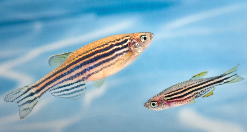
Dysferlin-null Zebrafish
Dysferlin null and other knock out zebrafish lines are available from Dr. Peter Currie at Monash University in Australia. These fish models were created using zinc finger technology from Sigma. Most of them have related phenotypes but they have not been characterized in detail yet, contact Dr. Currie (peter.currie@monash.edu) for more details.

Available Lines:
- Dysferlin
- Alpha-Dystrobrevin
- Alpha-Sarcoglycan
- ANHAK
- ANKAK2
- Beta-Parvin
- Collagen-6-alpha1
- FKRP
- FKTN
- Integrin-A7
- Myoferlin-1
- Myoferlin-2
- Otoferlin-1
- Otoferlin-2
- Sepn-1
More information on some knock out lines can be downloaded as an excel spreadsheet: Ferlin-Class-ZFN-Zebrafish-KOs.
Requests:
Because these fish lines are still unpublished, they are only available through a collaboration with Dr. Currie. These fish were made through a collaboration with Sigma and to use the fish, researchers must complete an MTA_SIGMA. Please email Dr. Currie at peter.currie@monash.edu for more information and to request the fish.
